
Unbiased headlines. Facts, not spin.
Every story is an unbiased news briefing written from 110+ sources across the spectrum — sources linked so you can verify it yourself.
 health
Notable (6/10)
Event date: September 16, 2026
health
Notable (6/10)
Event date: September 16, 2026
Scientists Identify Rare Brain Cells That Actively Switch the Cortex Into Sleep
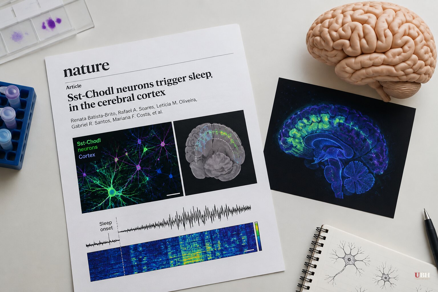
A Sleep Switch Hiding Inside the Cortex
Neuroscientists long treated the cerebral cortex as a passive rider during sleep, taking its cues from deep-brain structures like the hypothalamus and thalamus. A study published in Nature and led by Renata Batista-Brito, now an associate professor of neuroscience at the Icahn School of Medicine at Mount Sinai, shows that's wrong.
The research, conducted while Batista-Brito ran her lab at Albert Einstein College of Medicine, identified a rare group of cortical cells called Sst-Chodl neurons that can flip the brain into sleep mode by themselves. Flip the switch on these cells in a mouse, and the animal falls asleep, according to Geoffrey Terral, the study's lead author and a neuroscientist at Einstein.
"What our work shows is that the cortex can not only see this rhythm but also initiate it by itself, and this is sufficient to promote sleep," Terral said, according to Ars Technica.
The numbers involved are tiny. Sst-Chodl cells make up roughly one percent of the cortex's inhibitory neurons, according to Ars Technica, which works out to somewhere around one in every 500 to 1,000 cortical cells overall. Mount Sinai's own release puts the figure at approximately 0.2 percent of all cortical neurons. Either way, it's a needle-in-a-haystack population.
What makes them unusual isn't just their rarity. Most inhibitory neurons only talk to their immediate neighbors. Sst-Chodl cells do the opposite: they receive precise inputs, then broadcast that signal across the entire visual cortex and beyond, into areas tied to touch, hearing, and spatial memory, Batista-Brito told Ars Technica.
When the team activated these neurons in freely moving mice, slow-wave and REM sleep both increased, mice fell asleep faster, and they retreated to their nests during the day, according to NewsBeep. The effect held even during the nocturnal animals' normal waking hours. "We could have them sleep more during the time that they are awake than they usually sleep during the day," Batista-Brito said.
What Flips the Switch Is Still Unknown
The paper's co-author, Thomas Kilduff, director of SRI International's Center for Neuroscience, had previously shown that after sleep deprivation, these cells become the most active in the cortex, according to NewsBeep. This suggests Sst-Chodl neurons act as the cortex's own pressure gauge for accumulated fatigue, separate from the body's circadian clock.
But Batista-Brito's team still doesn't know what actually turns these cells on in the first place. The work so far was also confined to the visual cortex. "One caveat of our work is that it was done in the visual cortex," she said, per NewsBeep. Her lab is now repeating the anatomy work in the prefrontal cortex, predicting they'll find inputs from the hypothalamus and thalamus feeding into a sleep-specific version of the same circuit.
The cells are conserved across salamanders, reptiles, and mammals including humans, according to respiratory-therapy's coverage of the study. The researchers argue this makes them a plausible entry point for understanding the sleep disruption seen across many psychiatric illnesses.
A High-Risk Project the Grant System Almost Killed
Batista-Brito told Ars Technica that pinning down these neurons required years of building a labeling strategy specific enough to isolate a population this small, since targeting either of the two marker genes alone captures a much larger, unrelated group of cells. "If you're not really specific, the contaminants are going to be much more dominant than the specific cells," she said.
The project almost didn't happen. "I wrote a bunch of grants on these projects that were always rejected because it was too high-risk," Batista-Brito said. Federal and institutional science funding tends to favor safer, incremental proposals over long-shot ideas that might fail entirely, even when the long shot pays off.
Elsewhere in Brain Research This Month
Stanford Medicine researchers, in a study published online in Nature on September 16, transplanted lab-grown human cortical tissue, called organoids, into mice bred to lack most of their own cerebral cortex, according to Stanford Medicine's own release. Led by Sergiu Pasca, the team found the human tissue survived, grew, and wired itself into the mouse's brain and spinal cord. Researchers also spotted a nerve cell type never before seen in lab culture, previously observed only in autopsied human brains. Pasca said the model should accelerate research into schizophrenia, epilepsy, profound autism, and cerebral palsy.
Separately, neurologist David Perlmutter argued in an EpochTV interview covered by the Epoch Times that Alzheimer's research focuses too heavily on plaques, tangles, and inflammation while underweighting metabolic dysfunction. Perlmutter, a fellow of the American College of Nutrition, contends that fasting insulin, not the standard fasting glucose test, is a better early warning sign, with an ideal range of roughly 2 to 5 micro-international units per milliliter. That view is his own clinical perspective from an interview, not the result of a new peer-reviewed trial, and it sits alongside, not in place of, the plaque-and-tangle model that continues to drive most Alzheimer's research funding.
Back in the sleep lab, the open question remains what actually flips the Sst-Chodl switch. Batista-Brito's team says its next results, from the prefrontal cortex, should show whether the same tiny cell population runs the same trick brain-wide or whether each region built its own version of the circuit.
Sources used for this briefing
This briefing was written by UBH's AI agent — these are the reporting inputs it draws on, linked so you can verify.