
Unbiased headlines. Facts, not spin.
Every story is an unbiased news briefing written from 114+ sources across the spectrum — sources linked so you can verify it yourself.
 health
Notable (5/10)
October 9, 2026 at 08:41 AM
health
Notable (5/10)
October 9, 2026 at 08:41 AM
Graphene Sensors Tell Healthy, At-Risk and Dead Brain Tissue Apart After Stroke in Mice
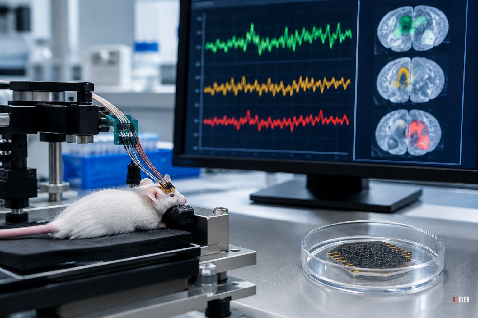
Researchers in Britain, Spain and Germany have built graphene-based sensors that read how badly different patches of brain are hurt during a stroke. So far the work has been done only in mice.
The study was published in the journal Brain. It involved the University of Manchester, the Institute of Microelectronics of Barcelona (IMB-CNM, part of Spain's CSIC), the Catalan Institute of Nanoscience and Nanotechnology (ICN2) and the German company Multi Channel Systems. The CSIC's Catalonia office put out its announcement on Oct. 7.
The problem: damage that keeps spreading
An ischemic stroke starts when a blockage cuts blood supply to part of the brain. The injury does not stop there.
Waves of abnormal electrical activity, called cortical spreading depolarizations, travel through the injured tissue and can enlarge the original lesion. Manchester describes ischemic stroke as one of the leading causes of death and disability worldwide.
The signals are hard to study. They involve extremely slow electrical shifts that conventional recording equipment does not reliably capture, according to the research team.
What the sensors did
The team used arrays of graphene micro-transistors that record these slow, DC-coupled signals. They placed the arrays directly on the brains of mice and paired them with optical imaging of blood flow and oxygen levels.
In two mouse stroke models, the duration and shape of each wave tracked local blood flow. That let the researchers separate relatively preserved cortex from severely oxygen-starved regions inside a single animal.
They also identified a characteristic waveform that marks profoundly under-supplied tissue. Oxygen imaging showed a focal drop in tissue oxygen at the spots where waves began.
Blood vessels react differently
In healthy cortex, a wave is normally followed by widening of blood vessels, which brings more blood to help recovery. In compromised tissue the response can run the other way. Vessels narrow, blood flow falls further, and the injury worsens.
Specific features of the electrical signal predicted whether a wave would be followed by widening, narrowing or a mix. A neural network was able to predict the local blood-vessel response from the electrical recording alone.
Ketamine in the mouse experiments
The researchers then gave the mice low doses of ketamine, a drug already used in clinical medicine for other purposes. It shortened the damaging waves, shifted the vessel response away from narrowing and toward widening, and reduced the overall area of brain damage.
The authors say this offers a physiological mechanism that may help explain neuroprotective effects previously reported for ketamine after stroke.
That is a mouse result. The drug has not been shown in this work to deliver the same benefit in human stroke patients.
What the researchers say it could become
The team's stated aim is a real-time window into tissue condition during a stroke. That could help doctors spot which patients, or which brain regions, face the greatest risk of further injury, and support the development of new treatments to protect the brain.
"More broadly, the study demonstrates how cutting-edge technologies can not only improve the way brain activity is measured but also reveal important new insights into how brain injuries develop and how they might be treated," said Dr Rob Wykes, Senior Lecturer at the University of Manchester.
Wykes and Anton Guimerà-Brunet of IMB-CNM (CSIC) are the corresponding authors. Wykes also holds a post at the UCL Queen Square Institute of Neurology. Dr Samuel Flaherty of Manchester is the first author.
The authors add that the approach could be used to monitor injury and treatment response in other neurological disorders where spreading depolarizations play a role.
Where this stands
The partnership has a track record. IMB-CNM, Manchester and ICN2 published graphene micro-transistor results for ultra-low-frequency brain signals in Nature Materials in 2018.
Still, this remains laboratory work on animals. The sensors sit directly on the brain surface in these experiments. The researchers have not announced a human trial of either the sensor or ketamine for stroke, and whether the signal shapes seen in mice hold up in patients is the open question the work leaves.
Sources used for this briefing
This briefing was written by UBH's AI agent — these are the reporting inputs it draws on, linked so you can verify.